 Hakan Gök
- Hernia Istanbul, Comprehensive Hernia Center, Istanbul, Türkiye
Introduction: The primary goal in incisional hernia repair is achieving primary fascial closure and reinforcing the area with a synthetic mesh. However, when Loss of Domain (LoD) is present, serious complications such as intra-abdominal hypertension (IAH) and abdominal compartment syndrome (ACS) may arise. Various strategies have been employed to overcome these challenges and increase the reduced intra-abdominal volume, including preoperative botulinum toxin (BTA) injection, progressive pneumoperitoneum (PPP), various component separation techniques, and their combinations. Intraoperative fascial traction (IFT) has recently been added to this armamentarium. The two cases presented here aim to demonstrate the potential benefits of this innovative technique and offer a different perspective to surgeons dealing with such challenging cases.
Presentation of Cases: The two patients presented here had previously undergone open umbilical hernia repair with mesh—one 17 years ago and the other 5 years ago—both of whom experienced recurrence and developed LoD over time. In both cases, IFT was successfully performed, resulting in an uneventful recovery.
Discussion: The repair of incisional hernias accompanied by LoD presents significant challenges. In managing these cases, it is essential not only to optimise the patient preoperatively but also to employ interventions aimed at increasing intra-abdominal volume. In recent years, the intraoperative fascial traction (IFT) technique has emerged as a valuable tool in complex incisional hernia repairs. This technique not only facilitates primary fascial closure but also significantly increases intra-abdominal volume, potentially reducing the risks associated with intra-abdominal hypertension and compartment syndrome.
Conclusion: IFT offers promising advantages in the repair of incisional hernias with LoD, as it addresses the dual challenge of achieving primary fascial closure and restoring intra-abdominal volume. The two cases presented highlight the potential of this innovative technique in achieving successful outcomes. However, further research and larger studies are needed to fully establish its efficacy and long-term benefits in this challenging patient population.
Introduction
The treatment of incisional hernias with an associated loss of domain (LoD) presents significant challenges. These patients often suffer from comorbidities such as diabetes, obesity, chronic obstructive pulmonary disease (COPD), and cardiac conditions, which individually or in combination can complicate hernia repair [1–5]. Preoperative optimisation, including weight reduction, diabetes control, smoking cessation, and respiratory physiotherapy, is crucial in improving outcomes [5–7].
The primary goal in hernia repair is to achieve a durable and recurrence-free outcome. In incisional hernias, this goal is best accomplished by ensuring the closure of the anterior fascia, followed by reinforcement with a mesh, which minimises complications and recurrence [8–10]. However, in cases where LoD is present, the situation becomes more complex [11].
LoD refers to a condition where a significant portion of intra-abdominal organs resides outside the abdominal cavity within the hernia sac, leading to a reduced intra-abdominal volume. Preoperative imaging with a CT scan and volumetric analysis are essential to assess the presence and extent of LoD [12, 13]. In these patients, the challenge lies in reintegrating the displaced organs into the original abdominal cavity without causing intra-abdominal hypertension (IAH), which can impair organ perfusion and pulmonary function or, in severe cases, lead to abdominal compartment syndrome (ACS) [14–18].
To address these challenges, various techniques have been developed to increase intra-abdominal volume. Preoperative interventions such as botulinum toxin A (BTA) injection, progressive pneumoperitoneum (PPP), and anterior or posterior component separation can be employed individually or in combination based on patient-specific needs [19–28].
In recent years, intraoperative fascial traction (IFT) has emerged as a promising technique for managing patients with large defects and LoD. This approach not only facilitates primary fascial closure but also contributes to increasing intra-abdominal volume, thereby mitigating the risks of postoperative complications [29–31].
In this report, we present two case studies of patients with incisional hernias and LoD, where IFT was successfully employed to enhance intra-abdominal volume and achieve primary fascial closure.
Case Reports
Case 1
A 49-year-old male patient underwent open umbilical hernia repair with mesh 17 years ago. The hernia recurred shortly after surgery and progressively enlarged over time. The patient had a BMI of 30, type 2 diabetes, and a history of smoking one pack of cigarettes per day. Physical examination revealed a large, irreducible recurrent umbilical hernia (Figure 1). CT imaging showed a 6 × 7 cm hernia defect and a large sac containing small bowel loops under the skin, resembling an hourglass appearance (Figure 2). The Tanaka Index was calculated at 30%.
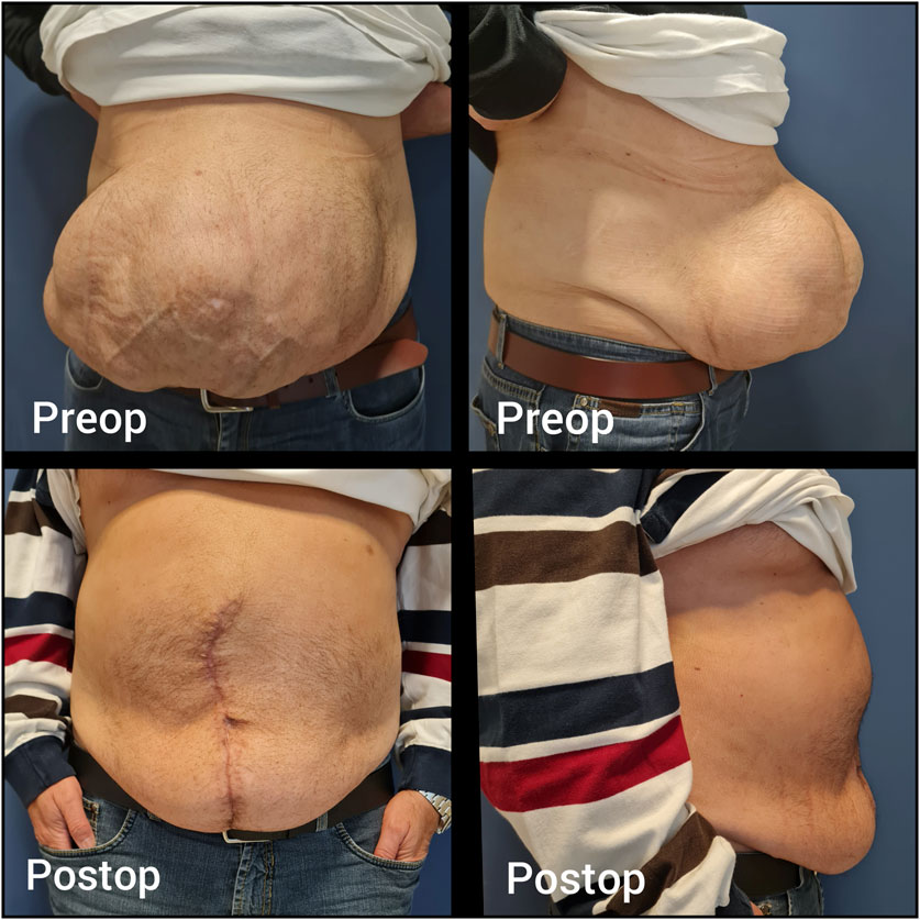
Figure 1. Case 1 - Preoperative and postoperative clinical appearance.
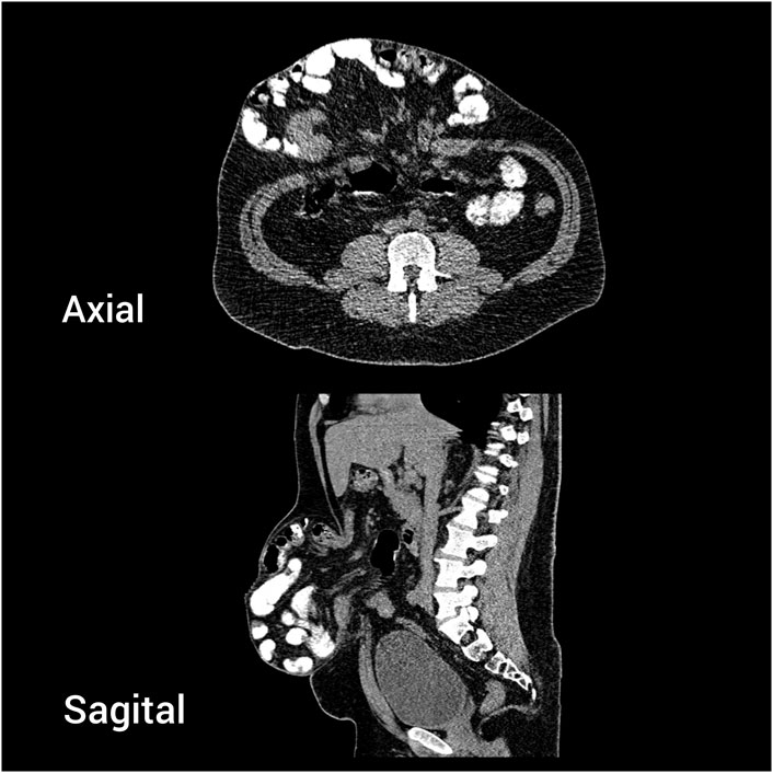
Figure 2. Case 1 - Preoperative CT scan. Axial and sagittal view.
One month prior to surgery, 200 U of botulinum toxin A (Botox®, Allergan Inc., United States) was injected bilaterally under ultrasound guidance into the external oblique (EO), internal oblique (IO), and transversus abdominis (TA) muscles. The patient was advised to quit smoking. However, a urinary nicotine test was not performed before surgery.
During surgery, complete adhesiolysis was performed, followed by a Rives-Stoppa dissection. A 15 × 20 cm medium-weight macroporous mesh was placed in the retrorectus space. IFT using the Fasciotens device (Fasciotens® GmbH, Germany) was applied, enabling successful primary fascial closure without pathological increases in intra-abdominal pressure (IAP). This was checked on the anaesthesia machine by monitoring the peak plateau pressures. The patient developed postoperative paralytic ileus, which resolved with non-operative operative treatment. The patient was discharged without any other complications on postoperative day 5. At the 1-year follow-up, no recurrence was observed (Figure 1).
Case 2
A 64-year-old male patient underwent open umbilical hernia repair with mesh 5 years ago. Recurrence occurred 2 months after surgery, and the hernia progressively enlarged over time. The patient had a BMI of 36.7 and well-controlled hypertension on medication. Physical examination revealed a large, irreducible recurrent umbilical hernia (Figure 3). CT imaging showed a 9 × 12 cm hernia defect and a large sac containing small bowel loops under the skin (Figure 4). The Tanaka Index was calculated at 51%.
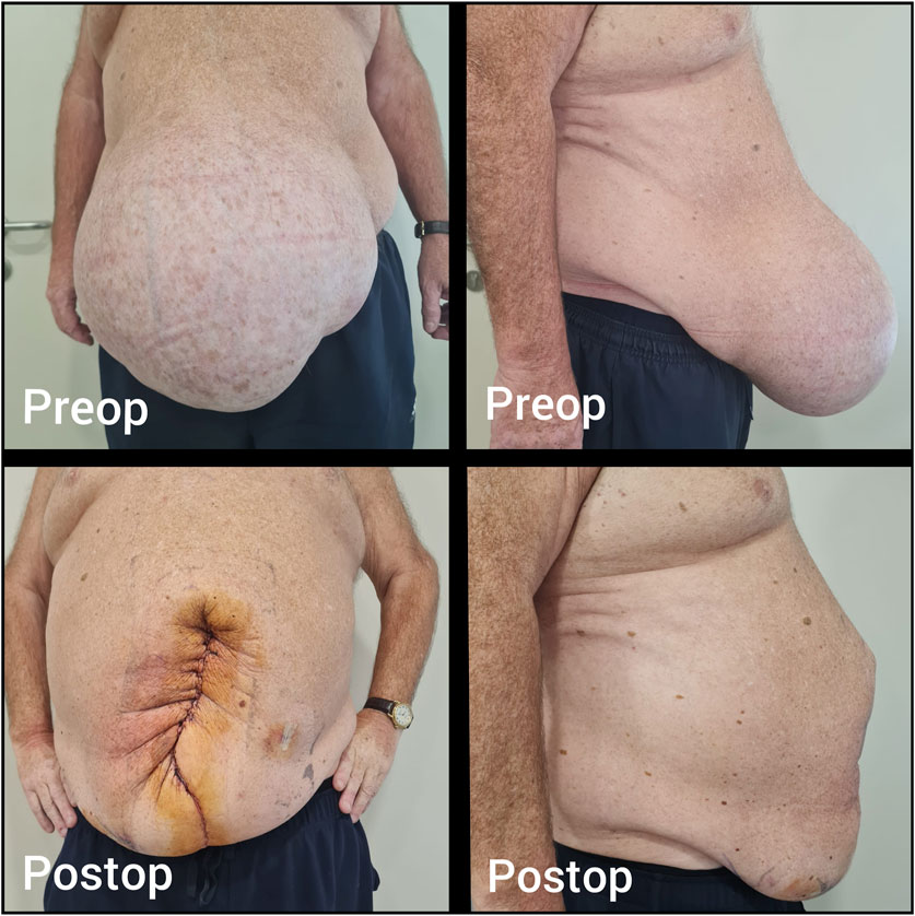
Figure 3. Case 2 - Preoperative and postoperative clinical appearance.
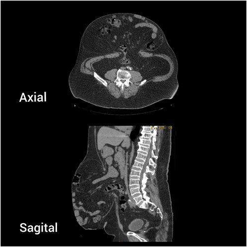
Figure 4. Case 2 - Preoperative CT scan. Axial and sagittal view.
One month prior to surgery, 200 U of botulinum toxin A (Botox®, Allergan Inc., United States) was injected bilaterally under ultrasound guidance into the EO and IO muscles.
During surgery, complete adhesiolysis was performed, followed by a Rives-Stoppa dissection. An 18 × 30 cm medium-weight macroporous mesh was placed in the retrorectus space. IFT using the Fasciotens device (Fasciotens® GmbH, Germany) was applied, allowing successful primary fascial closure without pathological increases in IAP. The patient was discharged without complications on postoperative day 1. At the time of writing, the patient was in the first postoperative week, with no complications or recurrence observed (Figure 3).
Discussion
Incisional hernias represent one of the most challenging aspects of abdominal wall hernias. They are not static; they grow over time and significantly impair quality of life [1, 2]. Their repair is complex, with recurrence rates reported as high as 5%–63% [1, 3, 10].
The primary method for reducing recurrence and complications in incisional hernia repair is achieving primary fascial closure [6, 8–10]. Additionally, reinforcement of the repair with a mesh placed in the retromuscular plane significantly reduces recurrence rates. However, in large hernias with LoD returning herniated organs to the abdominal cavity may lead to IAH, potentially resulting in ACS [14–18]. This pathological condition can impair respiratory function, compromise organ perfusion, and may lead to fatal outcomes.
To prevent this, various options are available to increase intra-abdominal volume. These include the use of BTA, which relaxes and elongates the external oblique (EO), internal oblique (IO), and transversus abdominis (TA) muscles, thus increasing abdominal volume [19–23] Although BTA provides a limited volume increase, it significantly contributes to achieving primary fascial closure by simplifying the complexity of the repair, it may downstage the procedure, reducing the need for anterior or posterior component separation (PCS) and transversus abdominis release (TAR), and allowing the repair to be completed with the Rives-Stoppa technique alone [9, 21]. BTA application has become nearly standard for midline hernias classified as EHS W3 (wider than 10 cm) [32]. In our two cases, we utilised BTA both to assist with primary fascial closure and increase intra-abdominal volume.
Another effective method for increasing intra-abdominal volume is the use of component separation techniques (CST). Two primary techniques are widely practiced today: Ramirez’s anterior component separation (ACS) [27] and Novitsky’s PCS with TAR [8]. Both techniques are effective in achieving primary fascial closure in midline hernias. However, they are technically demanding. While ACS carries risks of surgical site occurrences (SSO) such as hematoma, seroma, infection, and skin necrosis, PCS-TAR may result in seroma, infection, or challenging-to-repair EIT Ambivium hernias [10, 33–38]. Therefore, expertise in these techniques and careful case selection are essential.
Progressive pneumoperitoneum (PPP) is another effective method to increase intra-abdominal volume [24–26, 39]. In this technique, a catheter is placed into the abdominal cavity, and ambient air is gradually insufflated over 7–10 days preoperatively to expand the abdominal cavity. While effective, PPP is associated with significant complications such as patient intolerance, respiratory insufficiency, bowel injury, and thromboembolic events [26, 39, 40]. It is also time-consuming and costly.
Recently, the use of IFT has gained popularity, with multicenter studies reporting successful outcomes, particularly in achieving primary fascial closure [29–31]. This technique involves placing 12 traction sutures on the medial edge of each rectus muscle and applying a vertical force (12–20 kg) over 30 min, progressively medializing the rectus complex while also increasing intra-abdominal volume.
In our two cases with midline incisional hernias and LoD, we successfully utilised IFT. For the first case, where the defect was only 6 cm wide, one might argue that it could have been easily closed without requiring any additional intervention to achieve the goal. However, the focus was solely on increasing volume. In the second case, both volume increase and primary fascial closure were achieved without causing IAH.
During retrorectus dissection, when the posterior rectus sheath is separated from the muscle, the rectus muscles lose their cylindrical structure, elongate, and expand the retrorectus space. Therefore, as observed in the second case, the available space for mesh placement is greater than the preoperatively measured rectus widths on the CT scan. However, for a 9 cm defect, an 18 cm wide mesh may appear to provide insufficient overlap. In cases of large defects, there is an ongoing debate among hernia surgeons regarding the necessity of posterior component separation and TAR to achieve adequate mesh overlap. While some advocate for TAR, other centers have reported excellent outcomes without it, and both approaches have been supported by published data [8, 31, 39].
IFT is an easy-to-perform and effective technique. No significant complications specific to the technique or device have been reported to date [41, 42]. It can be combined with BTA, CST, and PPP techniques.
It is essential to recognise that no single technique can be universally applied to all hernias. Tailoring is key. Each case must be evaluated individually to determine the most suitable approach, utilizing one or a combination of the aforementioned techniques to optimise patient outcomes.
Limitations
This study presents the early outcomes of two cases, with one patient having a follow-up period of 1 year and the other only 1 week. Given the limited sample size and short follow-up duration, these findings cannot be generalised. Further studies with larger cohorts and long-term follow-up are needed to draw more definitive conclusions.
Conclusion
The management of incisional hernias, particularly those with large defects and LoD, requires a comprehensive and individualised approach. Achieving primary fascial closure remains the cornerstone of repair, but addressing the dual challenges of fascial closure and restoring intra-abdominal volume necessitates the integration of innovative techniques such as BTA, CST, PPP, and IFT.
IFT, in particular, offers promising advantages by effectively increasing intra-abdominal volume and facilitating primary fascial closure with minimal complications. The two cases presented underscore the potential of this innovative technique in achieving successful outcomes. By tailoring the surgical approach to the specific needs of each patient, combining one or more of these advanced techniques, surgeons can optimise outcomes, reduce recurrence rates, and improve overall patient quality of life.
However, further research and larger studies are essential to fully establish the long-term efficacy and benefits of IFT in this challenging patient population.
Data Availability Statement
The raw data supporting the conclusions of this article will be made available by the authors, without undue reservation.
Ethics Statement
Ethical approval was not required for the studies involving humans because the patients underwent a well-established surgical treatment. The studies were conducted in accordance with the local legislation and institutional requirements. The participants provided their written informed consent to participate in this study. Written informed consent was obtained from the individual(s) for the publication of any potentially identifiable images or data included in this article.
Author Contributions
HG conducted the primary data collection, analysis, and manuscript writing.
Funding
The author(s) declare that no financial support was received for the research, authorship, and/or publication of this article.
Conflict of Interest
The author declares that the research was conducted in the absence of any commercial or financial relationships that could be construed as a potential conflict of interest.
Generative AI Statement
The author(s) declare that no Generative AI was used in the creation of this manuscript.
Publisher’s Note
All claims expressed in this article are solely those of the authors and do not necessarily represent those of their affiliated organizations, or those of the publisher, the editors and the reviewers. Any product that may be evaluated in this article, or claim that may be made by its manufacturer, is not guaranteed or endorsed by the publisher.
Abbreviations
LoD, Loss of Domain; IFT, Intraoperative Fascial Traction; BTA, Botulinum Toxin A; CT, Computerised Tomography; PPP, Progressive Penumo Peritoneum; CST, Component Seperation Technique; ACS, Anterior Component Seperation; PCS, Posterior Component Seperation; TAR, Transeversus Abdominis Release; IAH, Intraabdominal Hypertension; COPD, Chronic Obstructive Pulmonary Disease; ACS, Abdominal Compartment Syndrome.
References
1. Hoffmann, H, Köckerling, F, Adolf, D, Mayer, F, Weyhe, D, Reinpold, W, et al. Analysis of 4,015 Recurrent Incisional Hernia Repairs from the Herniamed Registry: Risk Factors and Outcomes. Hernia (2021) 25(1):61–75. doi:10.1007/s10029-020-02263-x
2. van Ramshorst, GH, Eker, HH, Hop, WC, Jeekel, J, and Lange, JF. Impact of Incisional Hernia on Health-Related Quality of Life and Body Image: A Prospective Cohort Study. Am J Surg (2012) 204(2):144–50. doi:10.1016/j.amjsurg.2012.01.012
3. Bhardwaj, P, Huayllani, MT, Olson, MA, and Janis, JE. Year-Over-Year Ventral Hernia Recurrence Rates and Risk Factors. JAMA Surg (2024) 159(6):651–8. doi:10.1001/jamasurg.2024.0233
4. Sara, CG, Podda, M, Ribas, S, Montori, G, Botteri, E, Agresta, F, et al. What Defines an Incisional Hernia as ‘complex’: Results from a Delphi Consensus Endorsed by the European Hernia Society (EHS). Br J Surg (2024) 111(1):znad346. doi:10.1093/bjs/znad346
5. Sanders, DL, Pawlak, MM, Simons, MP, Aufenacker, T, Balla, A, Berger, C, et al. Midline Incisional Hernia Guidelines: The European Hernia Society. Br J Surg (2023) 110(12):1732–68. doi:10.1093/bjs/znad284
6. Ayuso, SA, Elhage, SA, Salvino, MJ, Sacco, JM, and Heniford, BT. State-of-the-art Abdominal Wall Reconstruction and Closure. Langenbecks Arch Surg (2023) 408(1):60. doi:10.1007/s00423-023-02811-w
7. Ayuso, SA, Elhage, SA, Fischer, JP, and Heniford, BT. The Role of Prehabilitation in Abdominal Wall Reconstruction: It Is More Than “Watch and Wait”. Ann Surg (2024) 5(2):e449. doi:10.1097/AS9.0000000000000449
8. Novitsky, YW, Elliott, HL, Orenstein, SB, and Rosen, MJ. Transversus Abdominis Muscle Release: A Novel Approach to Posterior Component Separation during Complex Abdominal Wall Reconstruction. Am J Surg (2012) 204(5):709–16. doi:10.1016/j.amjsurg.2012.02.008
9. van Rooijen, MMJ, Yurtkap, Y, Allaeys, M, Ibrahim, N, Berrevoet, F, and Lange, JF. Fascial Closure in Giant Ventral Hernias after Preoperative Botulinum Toxin a and Progressive Pneumoperitoneum: A Systematic Review and Meta-Analysis. Surgery (2021) 170(3):769–76. doi:10.1016/j.surg.2021.03.027
10. Maloney, SR, Schlosser, KA, Prasad, T, Kasten, KR, Gersin, KS, Colavita, PD, et al. Twelve Years of Component Separation Technique in Abdominal Wall Reconstruction. Surgery (2019) 166(4):435–44. doi:10.1016/j.surg.2019.05.043
11. Parker, SG, Halligan, S, Blackburn, S, Plumb, AAO, Archer, L, Mallett, S, et al. What Exactly Is Meant by “Loss of Domain” for Ventral Hernia? Systematic Review of Definitions. World J Surg (2019) 43(2):396–404. doi:10.1007/s00268-018-4783-7
12. Tanaka, EY, Yoo, JH, Rodrigues, AJ, Utiyama, EM, Birolini, D, and Rasslan, S. A Computerized Tomography Scan Method for Calculating the Hernia Sac and Abdominal Cavity Volume in Complex Large Incisional Hernia with Loss of Domain. Hernia (2010) 14(1):63–9. doi:10.1007/s10029-009-0560-8
13. Sabbagh, C, Dumont, F, Robert, B, Badaoui, R, Verhaeghe, P, and Regimbeau, JM. Peritoneal Volume Is Predictive of Tension-free Fascia Closure of Large Incisional Hernias with Loss of Domain: A Prospective Study. Hernia (2011) 15(5):559–65. doi:10.1007/s10029-011-0832-y
14. Petro, CC, Raigani, S, Fayezizadeh, M, Rowbottom, JR, Klick, JC, Prabhu, AS, et al. Permissible Intraabdominal Hypertension Following Complex Abdominal Wall Reconstruction. Plast Reconstr Surg (2015) 136(4):868–81. doi:10.1097/PRS.0000000000001621
15. Van Hoef, S, Dries, P, Allaeys, M, Eker, HH, and Berrevoet, F. Intra-abdominal Hypertension and Compartment Syndrome after Complex Hernia Repair. Hernia (2024) 28(3):701–9. doi:10.1007/s10029-024-02992-3
16. Quintela, C, Freire, L, Marrana, F, Barbosa, E1, Guerreiro, E, and Ferreira, FC. Quaternary Abdominal Compartment Syndrome in Complex Ventral Hernias. Int J Abdom Wall Hernia Surg (2021) 4(2):39–44. doi:10.4103/ijawhs.ijawhs_43_20
17. Kirkpatrick, AW, Nickerson, D, Roberts, DJ, Rosen, MJ, McBeth, PB, Petro, CC, et al. Intra-Abdominal Hypertension and Abdominal Compartment Syndrome after Abdominal Wall Reconstruction: Quaternary Syndromes? Scand J Surg (2017) 106(2):97–106. doi:10.1177/1457496916660036
18. Vidal, MG, Ruiz Weisser, J, Gonzalez, F, Toro, MA, Loudet, C, Balasini, C, et al. Incidence and Clinical Effects of Intra-abdominal Hypertension in Critically Ill Patients. Crit Care Med (2008) 36(6):1823–31. doi:10.1097/CCM.0b013e31817c7a4d
19. Ibarra-Hurtado, TR, Nuño-Guzmán, CM, Miranda-Díaz, AG, Troyo-Sanromán, R, Navarro-Ibarra, R, and Bravo-Cuéllar, L. Effect of Botulinum Toxin Type A in Lateral Abdominal Wall Muscles Thickness and Length of Patients with Midline Incisional Hernia Secondary to Open Abdomen Management. Hernia (2014) 18(5):647–52. doi:10.1007/s10029-014-1280-2
20. Motz, BM, Schlosser, KA, and Heniford, BT. Chemical Components Separation: Concepts, Evidence, and Outcomes. Plast Reconstr Surg (2018) 142(3 Suppl. l):58S–63S. doi:10.1097/PRS.0000000000004856
21. Niu, EF, Kozak, GM, McAuliffe, PB, Amro, C, Bascone, C, Honig, SE, et al. Preoperative Botulinum Toxin for Abdominal Wall Reconstruction in Massive Hernia Defects-A Propensity-Matched Analysis. Ann Plast Surg (2023) 90(6S Suppl. 5):S543–S546. doi:10.1097/SAP.0000000000003488
22. Soltanizadeh, S, Helgstrand, F, and Jorgensen, LN. Botulinum Toxin A as an Adjunct to Abdominal Wall Reconstruction for Incisional Hernia. Plast Reconstr Surg Glob Open (2017) 5(6):e1358. doi:10.1097/GOX.0000000000001358
23. Nielsen, MØ, Bjerg, J, Dorfelt, A, Jørgensen, LN, and Jensen, KK. Short-term Safety of Preoperative Administration of Botulinum Toxin A for the Treatment of Large Ventral Hernia with Loss of Domain. Hernia (2020) 24(2):295–9. doi:10.1007/s10029-019-01957-1
24. Moreno, IG. Chronic Eventrations and Large Hernias; Preoperative Treatment by Progressive Pneumoperitomeum; Original Procedure. Surgery (1947) 22(6):945–53.
25. Elstner, KE, Read, JW, Rodriguez-Acevedo, O, Ho-Shon, K, Magnussen, J, and Ibrahim, N. Preoperative Progressive Pneumoperitoneum Complementing Chemical Component Relaxation in Complex Ventral Hernia Repair. Surg Endosc (2017) 31(4):1914–1922. doi:10.1007/s00464-016-5194-1
26. Giuffrida, M, Biolchini, F, Capelli, P, Banchini, F, and Perrone, G. Botulinum Toxin and Progressive Pneumoperitoneum in Loss of Domain Ventral Hernias: A Systematic Review. J Abdom Wall Surg (2024) 3:12650. doi:10.3389/jaws.2024.12650
27. Ramirez, OM, Ruas, E, and Dellon, AL. Components Separation Method for Closure of Abdominal-Wall Defects: An Anatomic and Clinical Study. Plast Reconstr Surg (1990) 86(3):519–26. doi:10.1097/00006534-199009000-00023
28. De Luca, M, Medina Pedrique, M, Morejon Ruiz, S, Munoz-Rodriguez, JM, Robin Valle de Lersundi, A, Lopez-Monclus, J, et al. The Madrid Posterior Component Separation: An Anatomical Approach for Effective Reconstruction of Complex Midline Hernias. J Abdom Wall Surg (2024) 3:12928. doi:10.3389/jaws.2024.12928
29. Eucker, D, Zerz, A, and Steinemann, DC. Abdominal Wall Expanding System Obviates the Need for Lateral Release in Giant Incisional Hernia and Laparostoma. Surg Innov (2017) 24(5):455–61. doi:10.1177/1553350617718065
30. Niebuhr, H, Reinpold, W, Morgenroth, F, Berger, C, Dag, H, Wehrenberg, U, et al. Assessment of Myofascial Medialization Following Intraoperative Fascial Traction (IFT) in a Cadaveric Model. Hernia (2024) 28(4):1187–93. doi:10.1007/s10029-024-03003-1
31. Niebuhr, H, Aufenberg, T, Dag, H, Reinpold, W, Peiper, C, Schardey, HM, et al. Intraoperative Fascia Tension as an Alternative to Component Separation. A Prospective Observational Study. Front Surg (2021) 7:616669. doi:10.3389/fsurg.2020.616669
32. Muysoms, FE, Miserez, M, Berrevoet, F, Campanelli, G, Champault, GG, Chelala, E, et al. Classification of Primary and Incisional Abdominal Wall Hernias. Hernia (2009) 13(4):407–14. doi:10.1007/s10029-009-0518-x
33. Wegdam, JA, de Jong, DLC, de Vries Reilingh, TS, Schipper, EE, Bouvy, ND, and Nienhuijs, SW. Assessing Textbook Outcome after Implementation of Transversus Abdominis Release in a Regional Hospital. J Abdom Wall Surg (2022) 1:10517. doi:10.3389/jaws.2022.10517
34. Krpata, DM, Blatnik, JA, Novitsky, YW, and Rosen, MJ. Posterior and Open Anterior Components Separations: A Comparative Analysis. Am J Surg (2012) 203(3):318–22. discussion 322. doi:10.1016/j.amjsurg.2011.10.009
35. Oprea, V, Toma, M, Grad, O, Bucuri, C, Pavel, P, Chiorescu, S, et al. The Outcomes of Open Anterior Component Separation versus Posterior Component Separation with Transversus Abdominis Release for Complex Incisional Hernias: A Systematic Review and Meta-Analysis. Hernia (2023) 27(3):503–17. doi:10.1007/s10029-023-02745-8
36. Nielsen, MF, de Beaux, A, and Tulloh, B. Peritoneal Flap Hernioplasty for Reconstruction of Large Ventral Hernias: Long-Term Outcome in 251 Patients. World J Surg (2019) 43(9):2157–63. doi:10.1007/s00268-019-05011-0
37. Munoz-Rodriguez, JM, Lopez-Monclus, J, San Miguel Mendez, C, Perez-Flecha Gonzalez, M, Robin-Valle de Lersundi, A, Blázquez Hernando, LA, et al. Outcomes of Abdominal Wall Reconstruction in Patients with the Combination of Complex Midline and Lateral Incisional Hernias. Surgery (2020) 168(3):532–42. doi:10.1016/j.surg.2020.04.045
38. Vierstraete, M, Pereira Rodriguez, JA, Renard, Y, and Muysoms, F. EIT Ambivium, Linea Semilunaris, and Fulcrum Abdominalis. J Abdom Wall Surg (2023) 2:12217. doi:10.3389/jaws.2023.12217
39. Renard, Y, Lardière-Deguelte, S, de Mestier, L, Appere, F, Colosio, A, Kianmanesh, R, et al. Management of Large Incisional Hernias with Loss of Domain: A Prospective Series of Patients Prepared by Progressive Preoperative Pneumoperitoneum. Surgery (2016) 160(2):426–35. doi:10.1016/j.surg.2016.03.033
40. Bueno-Lledó, J, Martínez-Hoed, J, Bonafé-Diana, S, García-Pastor, P, Torregrosa-Gallud, A, Pareja-Ibars, V, et al. Complications Related to the Prehabilitation with Preoperative Pneumoperitoneum in Loss of Domain Hernias: Our Experience in 180 Consecutive Cases. Hernia (2024) 28(5):1591–8. doi:10.1007/s10029-023-02836-6
41. Eucker, D, Rüedi, N, Luedtke, C, Stern, O, Niebuhr, H, Zerz, A, et al. Abdominal Wall Expanding System. Intraoperative Abdominal Wall Expansion as a Technique to Repair Giant Incisional Hernia and Laparostoma. New and Long-Term Results from a Three-Center Feasibility Study. Surg Innov (2022) 29(2):169–82. doi:10.1177/15533506211041477
Keywords: incisional hernia, loss of domain, intraoperative fascial traction, abdominal compartment syndrome, complex hernia
Citation: Gök H (2025) Case Report: Intraoperative Fascial Traction for Increasing Intra-Abdominal Volume in Loss-of-Domain Incisional Hernias: A Report of Two Cases. J. Abdom. Wall Surg. 4:14283. doi: 10.3389/jaws.2025.14283
Received: 31 December 2024; Accepted: 28 February 2025;
Published: 14 March 2025.
Copyright © 2025 Gök. This is an open-access article distributed under the terms of the Creative Commons Attribution License (CC BY). The use, distribution or reproduction in other forums is permitted, provided the original author(s) and the copyright owner(s) are credited and that the original publication in this journal is cited, in accordance with accepted academic practice. No use, distribution or reproduction is permitted which does not comply with these terms.
*Correspondence: Hakan Gök, aGFrZ29rQGdtYWlsLmNvbQ==