Abstract
Skin aging is a multifaceted process influenced by genetic, environmental, and immunological factors, resulting in visible changes such as wrinkles, pigmentation, and alterations in skin texture. Despite its high relevance, there is a paucity of large-scale studies focusing on visible age-related changes in facial skin among Japanese populations, particularly by region. This study aims to address these gaps by examining age- and sex-specific skin changes in a large cohort of 2,543 Japanese subjects using advanced skin imaging techniques. The study included subjects aged 17–71 years who provided informed consent. The imaging system captures facial images using standard light, UV light, and two types of polarized light, applying deep learning techniques to analyze various skin parameters. Statistical analysis was performed to evaluate correlations between age, gender, and skin characteristics. Results indicated significant correlations between age and skin color across all domains, with large pores on the nose showing the highest correlation with age. Sebum and porphyrin levels exhibited a decreasing trend with age, though correlation coefficients were low. Cheek gloss, both in area and color, showed relatively high correlation coefficients. Pigmentation-related items, such as spots and melanin, demonstrated significant age correlations, particularly in areas like the corners of the eyes, under the eyes, and cheeks. Wrinkles and fine wrinkles correlated with age in various regions, though not on the forehead. The findings highlight the importance of understanding regional and demographic variations in skin aging to develop effective anti-aging treatments.
Introduction
Skin aging is a multifaceted process influenced by genetic, environmental, and immunological factors [1, 2]. This complex interplay results in visible changes such as wrinkles, pigmentation, and alterations in skin texture. Understanding these changes is crucial for developing effective anti-aging treatments. Despite the high relevance, there is a paucity of large-scale studies focusing on visible age-related changes in facial skin among Japanese populations, particularly by region. Furthermore, evidence on the specific manifestations of skin aging remains sparse. The manifestations of skin aging vary significantly by anatomical region and ethnicity, with different populations showing distinct patterns of aging [3–5]. This study aims to address these gaps by examining age- and sex-specific skin changes in a large cohort of 2,543 Japanese subjects using a sophisticated skin analyzer.
Materials and methods
Subjects
This study included patients who underwent skin analysis using NeoVoir® (C-Lab Co., Ltd., Tokyo, Japan) at a single institution in Japan between February 2021 and March 2022. Subjects aged 17–71 years who provided informed consent were included, with the exclusion of those diagnosed with skin disease or who were pregnant or lactating. A total of 2,543 Japanese individuals (2,439 females and 104 males) participated in the study. The research protocol was approved by the Ethics Committee of the University of Tokyo School of Medicine (approval number: 0695) and adhered to the principles of the Declaration of Helsinki.
Research equipment
NeoVoir® is a device comprising a skin imaging machine and an analysis application. The machine captures facial images using four types of light: standard light, UV light, and two types of polarized light. The brightness during photo shooting was adjusted using the analysis application to ensure consistent conditions, and the focus was also standardized by photographing a chart included with the equipment. The analysis application was developed using data from 3,000 Korean individuals, and by referring to this data again, the program can be validated.
Region setting
Deep learning techniques are applied to these images to automatically set a total of 10 locations: forehead, dorsum of the nose, right and left eye corners, right and left under the eyes, right and left cheeks, and right and left nasolabial folds. A total of 20 items are detected for the above 10 locations, including the number of large/medium/small pores, wrinkles/fine wrinkles, skin color (R/G/B values and their average value), gloss area and color, redness, pigment changes other than redness, large/medium/small melanin, sebum, and porphyrin levels. These results are output as numeric values.
Principles and definitions
The device captures images using standard light, UV light, and two types of polarized light. The deep learning technique is applied to the acquired image, and the device automatically delineates the following regions: forehead, dorsum of the nose, outer corner of the eyes (right and left), under the eyes (right and left), cheeks (right and left), and nasolabial folds (right and left). In photographs taken with polarized light, the red components are recognized as redness, and areas different from normal skin color, excluding redness, are identified as spots.
The Shadow Method was used to assess pores and wrinkles. This method measures the shadow area by indirectly illuminating both sides and top and bottom of the brow with a general light source. Using this method, only the red (R) value is measured by excluding the green (G) and blue (B) values associated with the black color from the RGB. Since the black color is not measured, eyebrows and eyelashes are excluded, and shadows caused by pores can be identified by color. The number of shadows was then measured and defined as pores. Using the same Shadow Method, shadows thought to be caused by wrinkles were also measured and defined as wrinkles. By capturing images with horizontally polarized light that does not suppress light reflection, shadows not visible with ordinary light sources can be detected. Shadows considered to be caused by fine wrinkles were measured and defined as fine wrinkles.
Colors are represented by RGB values for each region photographed in standard light. The values for red, green, and blue in the RGB color model were used. In horizontally polarized light imaging, areas reflecting light and appearing white were identified as gloss, with their area and RGB average values calculated as color.
Melanin was defined as the dark area measured using only the shadow tone from the UV light source, where melanin can be easily detected. For pores and melanin, measurements were divided by size, with 20–70 pixels as small (S), 71 to 120 pixels as medium (M), and 121 pixels and larger as large (L). Sebum was identified by extracting only white and blue fluorescence using the high-light tone from the UV light source. Among the extracted sebum, only orange and red were extracted and designated as porphyrin.
Measurement
Before measurements, subjects were cleansed and kept at rest in an environment of 20°C–24°C and 48%–50% relative humidity for 30 min. Various measurements were made using photographs from the frontal view, based on the machine’s program. Subjects were photographed with their hair pulled back, attaching their forehead and lower jaw to the fixing device of the imaging equipment.
Statistical analysis
Numerical data were exported from NeoVoir® as CSV files and analyzed using Python 3.8. Average values were calculated for symmetrical areas (corners of the eyes, under the eyes, cheeks, and nasolabial folds). The forehead and nose were added to these, making a total of 6 areas and 106 items. In the figures, for simplicity, the average value of the outer corners of the eyes is labeled as “eye corners,” the average value of under the eyes is labeled as “under-eye,” and the dorsum of the nose is labeled as “nose.” The relationship to gender and age was then examined. To examine gender differences, normality was first assessed, and based on the results, differences between males and females were tested using the t-test or the Mann-Whitney U test. Due to the small number of male subjects, age-related examinations and correlations were conducted using only the female data. For age-related examination and correlations between measurement items, normality was first assessed, and correlations were examined using Pearson’s correlation coefficient for normally distributed items and Spearman’s rank correlation coefficient for non-normally distributed items. For sebum and porphyrin levels, subjects were divided into 10-year age groups, and the mean value at each age group was calculated, followed by one-way ANOVA.
Results
Subject background
The mean age of the subjects was 35.8 years, with a median age of 34 years. The study included a predominantly female cohort, with 2,439 female and 104 male subjects (Figure 1).
FIGURE 1
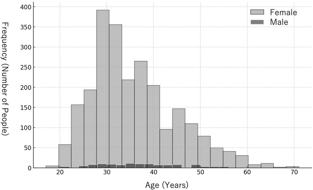
Age distribution of participants by gender. The histogram illustrates the distribution of ages for female (light gray) and male (black) participants.
Examination of skin changes associated with gender
Significant correlations were found in 86 items. The top 10 items with statistically significant differences were identified, and their distribution between males and females was visualized using box-and-whisker plots (Figure 2). The top 10 items were all related to color.
FIGURE 2
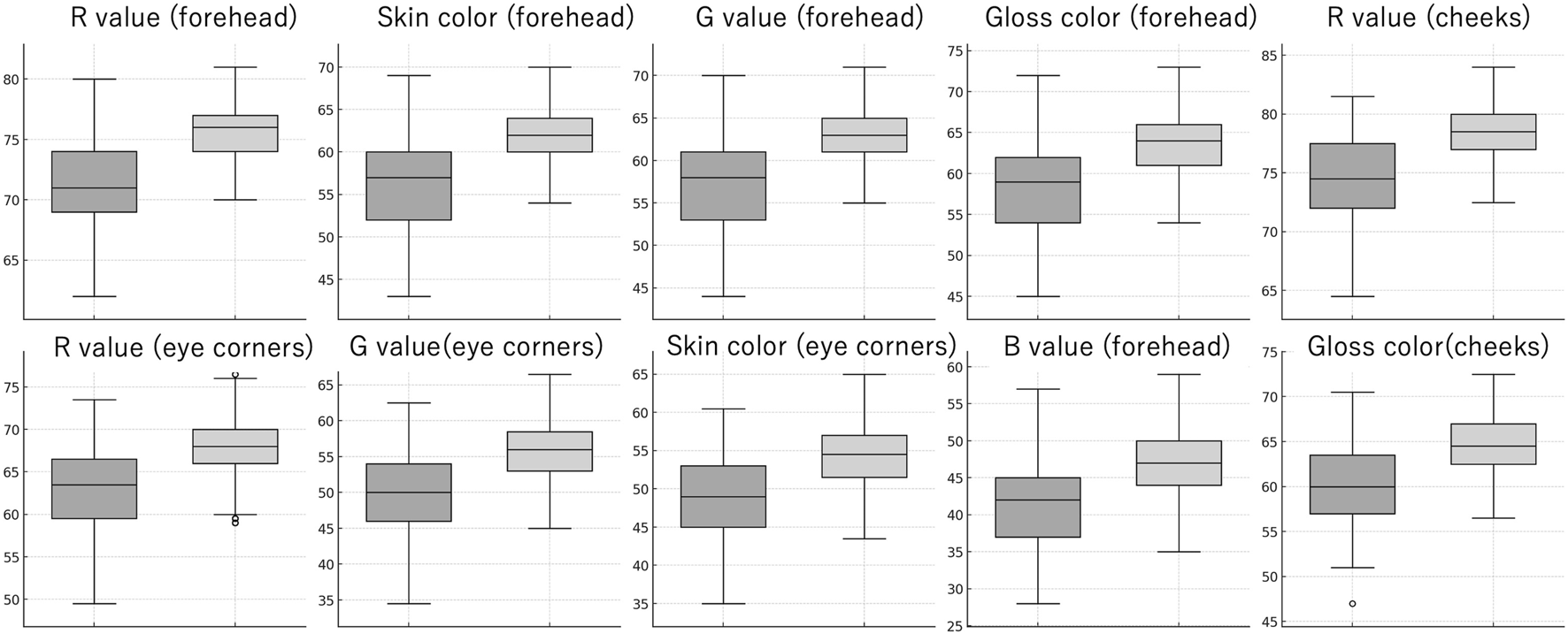
Box plots showing the top 10 parameters that have the highest correlation with gender. Each pair of box plots compares male (left, darker box) and female (right, lighter box) subjects. The box plots represent the median, interquartile range (IQR), and the full range of the data, with outliers indicated by individual points.
Examination of skin changes with age
Items related to skin color (skin color and R, G, B values) showed correlations with age across all domains. The top 10 items with statistically significant differences were identified and visualized using scatter plots (Figure 3). The degree of correlation varied by area and pore size, with the number of large pores on the nose showing the highest correlation with age. The top 10 items with statistically significant differences were identified and visualized using scatter plots (Figure 4). Sebum levels showed a decreasing trend with age on the forehead, nose, under the eyes, and cheeks, though the correlation coefficients were −0.144, −0.156, −0.064, and −0.114, respectively, indicating low correlations. Porphyrin levels also showed a decreasing trend with age on the forehead, nose, under the eyes, and cheeks, with correlation coefficients of −0.099, −0.115, −0.070, and −0.091, respectively, also indicating low correlations. ANOVA analysis for each age group revealed that both sebum and porphyrin levels decreased with age.
FIGURE 3
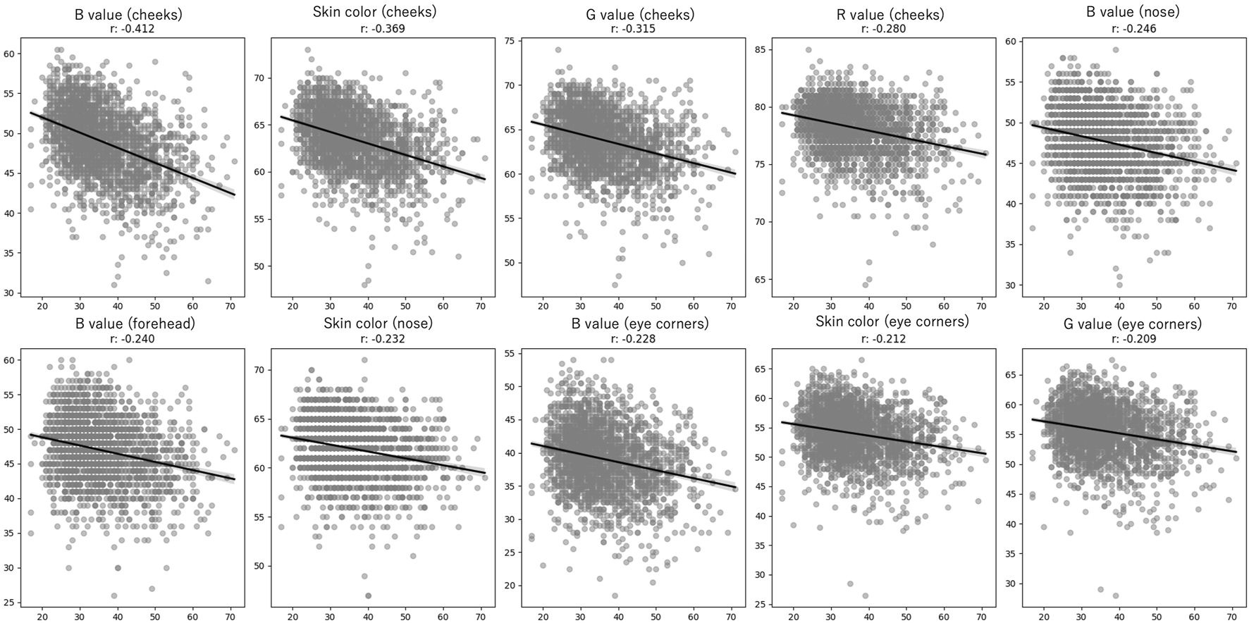
Correlation between age and skin color metrics in female subjects. The scatter plots depict the relationship between age and the top 10 parameters related to skin color with the strongest absolute correlation with age. Each subplot features a regression line (black) and the corresponding correlation coefficient (r) to indicate the strength of association between age and the pore variables. Individual measurements are shown as gray scatter points.
FIGURE 4
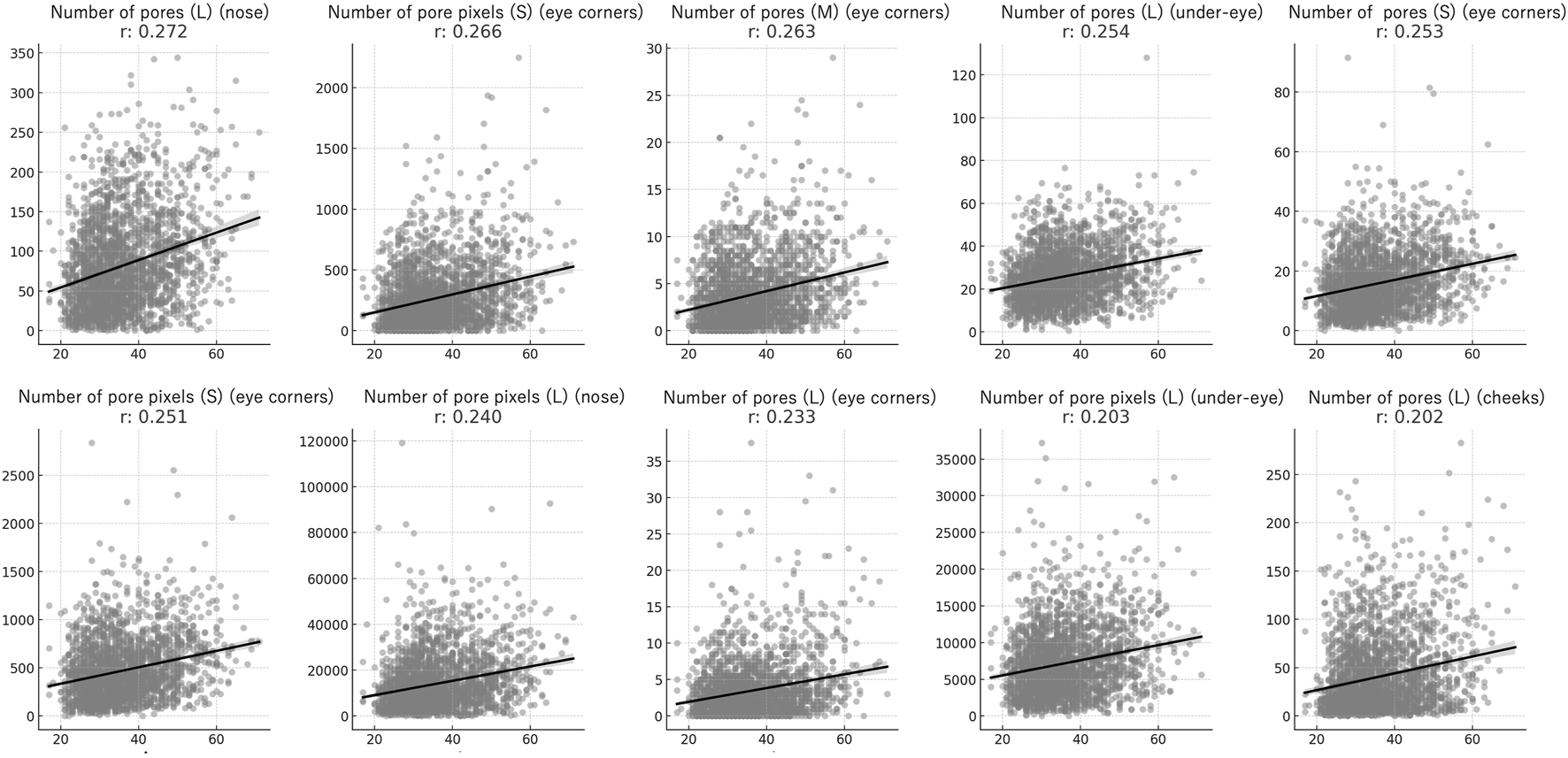
Correlation between age and pore metrics in female subjects. The scatter plots depict the relationship between age and the top 10 pore-related parameters with the strongest absolute correlation with age. Each subplot features a regression line (black) and the corresponding correlation coefficient (r) to indicate the strength of association between age and the pore variables. I Individual measurements are shown as gray scatter points.
For cheek gloss, both area and color had relatively high correlation coefficients (Figure 5). Redness did not show a significant correlation with age in any area. Spots and melanin, items related to pigmentation, showed correlations with age across all domains, with higher correlation coefficients at the corners of the eyes, under the eyes, and on the cheeks. The top 10 items with statistically significant differences in spots and melanin were identified and visualized using scatter plots (Figure 6). Wrinkles and fine wrinkles showed little correlation on the forehead but were correlated with age in other areas (Figure 7).
FIGURE 5
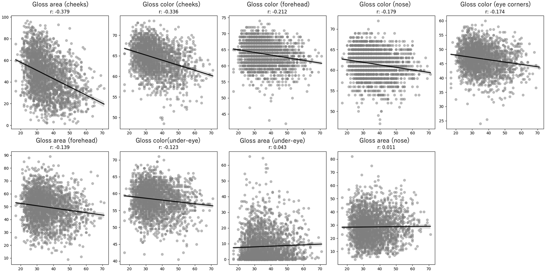
Correlation between age and skin glossiness metrics in female subjects. The scatter plots illustrate the relationship between age and the top 10 glossiness-related parameters that show the strongest absolute correlation with age. Each subplot features a regression line (black) and the corresponding correlation coefficient (r) to quantify the strength of association between age and glossiness variables. Individual measurements are shown as gray scatter points.
FIGURE 6
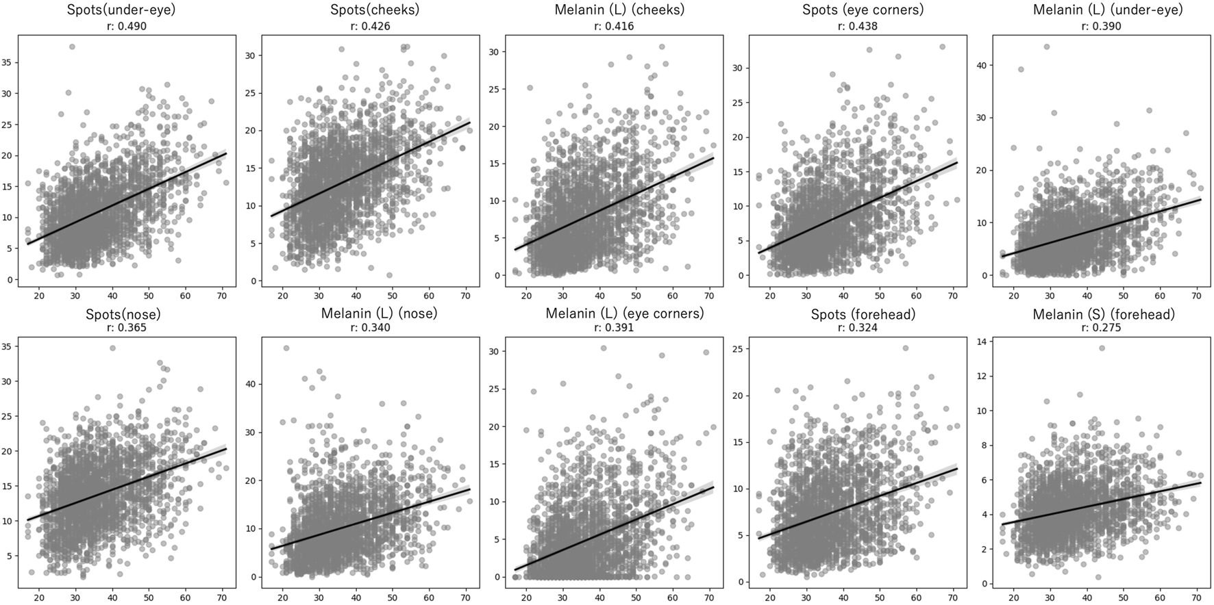
Correlation between age and pigmentation metrics in female subjects. The scatter plots represent the relationship between age and the top 10 pigmentation-related parameters with the highest absolute correlation with age. Each subplot includes a regression line (black) and the corresponding correlation coefficient (r) to indicate the strength of association between age and pigmentation variables. Individual measurements are shown as gray scatter points.
FIGURE 7
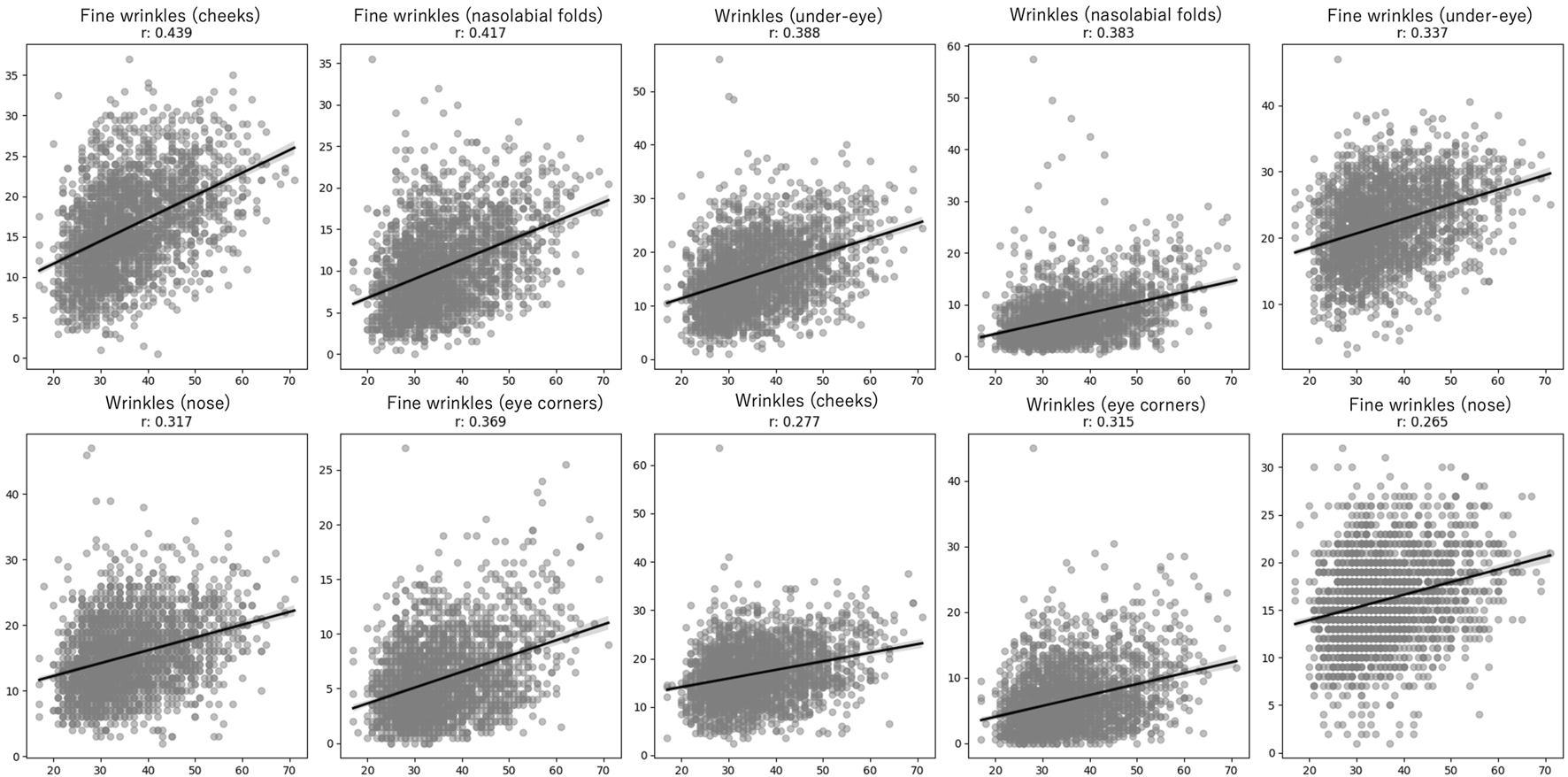
Correlation between age and wrinkle and fine wrinkle in female subjects. The scatter plots depict the relationship between age and the top 10 wrinkle-related parameters that show the highest absolute correlation with age. Each subplot includes a regression line (black) with corresponding Pearson or Spearman correlation coefficients (r) to quantify the strength of association between age and the respective wrinkle parameter. Individual measurements are shown as gray scatter points.
Discussion
The relationship between gender and skin changes has been extensively studied in various populations. Li et al. reported that women tend to have lighter skin color than men in exposed areas in a Chinese cohort [6]. Rahrovan et al. found that parameters such as skin water content, TEWL (transepidermal water loss), sebum content, capillary density, pigmentation, and thickness were generally higher in males [7]. Consistent with these findings, our study identified significant gender differences in skin color, particularly in exposed areas such as the forehead, cheeks, and corners of the eyes. In RGB values, black is represented by (0, 0, 0) and white by (255, 255, and 255), indicating that lighter colors have higher values. Our study found that women had higher RGB values, corresponding to lighter skin color.
Regarding the relationship between skin color and age, Kikuchi et al. observed that the b* value (yellowness) of the cheeks significantly increased with age in Japanese women, while the a* (redness) and L* (lightness) values did not show significant changes. This change was not observed in Caucasians [8]. Ogura et al. conducted skin biopsies on seven Japanese subjects and found that the b* value in the dermis increased in exposed areas compared to non-exposed areas, which they attributed to the carbonylation of matrix proteins [9]. In our study, B and G values, as well as the average RGB values, decreased with age. Although previous studies used L*a*b* color space to evaluate skin color, and our study used RGB color space, making direct comparisons difficult, the trends observed were consistent. The decrease in B and G values with age suggests that skin color becomes more yellow and less blue, which aligns with previous findings.
Flament et al. reported that pore size on the cheeks increased significantly with age in Indian women [10]. In Japanese women aged 18 to 29, pore size was initially small but increased with age, reaching a plateau after the age of 40. In our study, the number of large pores on the nose increased with age, and there was a corresponding increase in pixel count. Further studies on pores are needed to confirm these findings.
Sebum secretion is low in childhood, begins to increase during mid-to late childhood under the influence of androgens, and continues to rise until the late teens. It then remains relatively stable until later in life. In women, sebum secretion gradually decreases after menopause and does not change significantly after age 70. The most likely explanation for the age-related decrease in sebaceous gland secretion in both men and women is the simultaneous decrease in endogenous androgen production [11]. In this study, measurements were conducted 30 min after face washing due to time constraints. Liu et al. stated that evaluation 3 h after face washing is generally recommended and reported that sebum levels recover by 70%–90% within 1 h after sebum removal [12]. In this study, the short time elapsed after face washing may not have reflected the actual sebum secretion, which could explain the low correlation coefficient. Our study also observed a decrease in sebum with age in seborrheic areas such as the forehead and nose. There was a decreasing trend in sebum under the eyes and on the cheeks, although the correlation was weak, and no significant correlation was observed in the corners of the eyes. Porphyrin production by Cutibacterium acnes is well known and is associated with the severity of acne. Wu et al. reported that the amount of porphyrins on the face of Japanese individuals peaked in their 30 s and then decreased with age [13]. Our study found a similar decrease in porphyrin levels with age. In the NeoVoir® analysis application, sebum with orange or red components is recognized as porphyrin. Therefore, due to the limited amount of sebum resulting from the short time elapsed after washing, it is possible that the program could not detect sufficient porphyrin, leading to a low correlation coefficient. As such, differences in measurement methods and conditions may lead to variations in results, and further validation is needed.
Masuda et al. found that cheek gloss correlated with age [14]. In our study, no significant correlation was observed between the area of gloss under the nose and eyes and age. The relationship between skin reflectance and skin properties remains unclear, and further studies are needed to explore the biological changes associated with skin reflectance.
Our findings on redness were consistent with previous studies that found no relationship between redness and age [15, 16].
Regarding pigmentation, spots (areas different from normal skin color captured by polarized light, excluding redness) showed a stronger correlation with age than melanin. Ultraviolet light is known to enhance pigmented spots by being absorbed by melanin pigment [17]. Ultraviolet imaging, a photographic technique using only UV light, is a minimally invasive and accurate means of detecting pigmentary abnormalities [18]. Both spots and melanin showed high correlations with age, particularly in photoaged areas such as under the eyes, cheeks, and outer corners of the eyes. Larger melanin spots showed higher correlations with age, although small melanin spots on the forehead were most strongly correlated with age.
Fine wrinkles, detectable only by polarized light and not by ordinary light in the cheeks, outer corners of the eyes, and nasolabial folds, showed higher correlations with age. Although many studies have indicated that forehead wrinkles increase with age, our study did not find a significant correlation [3]. This suggests that the characteristics of wrinkles, such as size and depth, may vary by region, requiring different evaluation methods.
There are several limitations to this study. First, the gender distribution of the subjects was heavily skewed, with a majority being female and only 104 male subjects. This imbalance may have limited our ability to fully evaluate gender differences. Future studies should aim to include a more balanced gender distribution to better understand these differences. Second, the cross-sectional nature of the study limits the ability to draw conclusions about causal relationships between age and skin characteristics. Longitudinal studies are needed to confirm these findings and understand the progression of skin changes over time. Third, the study was conducted at a single institution, which may limit the generalizability of the results to other populations or regions. Multi-center studies with diverse populations are recommended to validate these results.
Conclusion
This study analyzed the results of the imaging system measurements on 2,543 Japanese subjects, identifying key factors related to gender and age that may enhance the efficiency of aging assessments and rejuvenation therapies. Given the significant differences in aging manifestations across races, measurement methodologies, and regions, further accumulation of data specific to the Japanese population is essential for developing more precise and effective interventions.
Statements
Data availability statement
The data that support the findings of this study are available from the corresponding author upon reasonable request. Requests to access these datasets should be directed to AY, ayuyoshi@me.com.
Ethics statement
The studies involving humans were approved by the Ethics Committee of the University of Tokyo Hospital. The studies were conducted in accordance with the local legislation and institutional requirements. Written informed consent for participation in this study was provided by the participants’ legal guardians/next of kin.
Author contributions
All authors participated in the design, interpretation of the studies and analysis of the data and review of the manuscript; JO, SS, and AY performed data collection, YO and JO were responsible for the statistical analysis and data interpretation, YO wrote the initial draft of the manuscript, JO and AY reviewed and edited the manuscript for important intellectual content. All authors contributed to the article and approved the submitted version.
Funding
The author(s) declare that no financial support was received for the research, authorship, and/or publication of this article.
Conflict of interest
AY belongs to the Social Cooperation Program, Department of Clinical Cannabinoid Research, supported by Japan Cosmetic Association and Japan Federation of Medium&Small Enterprise Organizations.
The remaining authors declare that the research was conducted in the absence of any commercial or financial relationships that could be construed as a potential conflict of interest.
References
1.
Ok SC . Insights into the anti-aging prevention and diagnostic medicine and healthcare. Diagnostics (Basel) (2022) 12(4):819. 10.3390/diagnostics12040819
2.
He X Gao X Xie W . Research progress in skin aging and immunity. Int J Mol Sci (2024) 25(7):4101. 10.3390/ijms25074101
3.
Ezure T Amano S . The severity of wrinkling at the forehead is related to the degree of ptosis of the upper eyelid. Skin Res Technol (2010) 16(2):202–9. 10.1111/j.1600-0846.2010.00427.x
4.
Toda K Pathak MA Parrish JA Fitzpatrick TB Quevedo WC Jr . Alteration of racial differences in melanosome distribution in human epidermis after exposure to ultraviolet light. Nat New Biol (1972) 236(66):143–5. 10.1038/newbio236143a0
5.
Tsukahara K Sugata K Osanai O Ohuchi A Miyauchi Y Takizawa M et al Comparison of age-related changes in facial wrinkles and sagging in the skin of Japanese, Chinese and Thai women. J Dermatol Sci (2007) 47(1):19–28. 10.1016/j.jdermsci.2007.03.007
6.
Li X Galzote C Yan X Li L Wang X . Characterization of Chinese body skin through in vivo instrument assessments, visual evaluations, and questionnaire: influences of body area, inter-generation, season, sex, and skin care habits. Skin Res Technol (2014) 20(1):14–22. 10.1111/srt.12076
7.
Rahrovan S Fanian F Mehryan P Humbert P Firooz A . Male versus female skin: what dermatologists and cosmeticians should know. Int J Womens Dermatol (2018) 4(3):122–30. 10.1016/j.ijwd.2018.03.002
8.
Kikuchi K . Influence of environmental stress on skin tone, color and melanogenesis in Japanese skin. Int J Cosmet Sci (2005) 27(1):52–4. 10.1111/j.1467-2494.2004.00254_9.x
9.
Ogura Y Kuwahara T Akiyama M Tajima S Hattori K Okamoto K et al Dermal carbonyl modification is related to the yellowish color change of photo-aged Japanese facial skin. J Dermatol Sci (2011) 64(1):45–52. 10.1016/j.jdermsci.2011.06.015
10.
Flament F Francois G Qiu H Ye C Hanaya T Batisse D et al Facial skin pores: a multiethnic study. Clin Cosmet Investig Dermatol (2015) 8:85–93. 10.2147/CCID.S74401
11.
Pochi PE Strauss JS Downing DT . Age-related changes in sebaceous gland activity. J Invest Dermatol (1979) 73(1):108–11. 10.1111/1523-1747.ep12532792
12.
Liu Y Jiang W Tang Y Zhang Q Zhen Y Wang X et al An optimal method for quantifying the facial sebum level and characterizing facial sebum features. Skin Res Technol (2023) 29(9):e13454. 10.1111/srt.13454
13.
Wu Y Akimoto M Igarashi H Shibagaki Y Tanaka T . Quantitative assessment of age-dependent changes in porphyrins from fluorescence images of ultraviolet photography by image processing. Photodiagnosis Photodyn Ther (2021) 35:102388. 10.1016/j.pdpdt.2021.102388
14.
Masuda Y Yagi E Oguri M Kuwahara T . Development of a quantitative method for evaluation of skin radiance and its relationship with skin surface topography. J Soc Cosmet Chem (2017) 51:211–8. 10.5107/sccj.51.211
15.
Lee E Cho C Ha J . Biophysical properties of redness-prone skin in Korean women. J Cosmet Dermatol (2022) 21(9):4035–41. 10.1111/jocd.14722
16.
Kikuchi K Masuda Y Yamashita T Kawai E Hirao T . Image analysis of skin color heterogeneity focusing on skin chromophores and the age-related changes in facial skin. Skin Res Technol (2015) 21(2):175–83. 10.1111/srt.12174
17.
Brenner M Hearing VJ . The protective role of melanin against UV damage in human skin. Photochem Photobiol (2008) 84(3):539–49. 10.1111/j.1751-1097.2007.00226.x
18.
Kojima K Shido K Tamiya G Yamasaki K Kinoshita K Aiba S . Facial UV photo imaging for skin pigmentation assessment using conditional generative adversarial networks. Sci Rep (2021) 11(1):1213. 10.1038/s41598-020-79995-4
Summary
Keywords
skin aging, facial analysis, sebum, wrinkles, pigmentation
Citation
Osuji Y, Omatsu J, Sato S and Yoshizaki A (2024) Age and gender differences in skin characteristics: a study of 2543 Japanese individuals using advanced skin imaging techniques. J. Cutan. Immunol. Allergy 7:13561. doi: 10.3389/jcia.2024.13561
Received
20 July 2024
Accepted
27 September 2024
Published
07 November 2024
Volume
7 - 2024
Updates
Copyright
© 2024 Osuji, Omatsu, Sato and Yoshizaki.
This is an open-access article distributed under the terms of the Creative Commons Attribution License (CC BY). The use, distribution or reproduction in other forums is permitted, provided the original author(s) and the copyright owner(s) are credited and that the original publication in this journal is cited, in accordance with accepted academic practice. No use, distribution or reproduction is permitted which does not comply with these terms.
*Correspondence: Ayumi Yoshizaki, ayuyoshi@me.com
Disclaimer
All claims expressed in this article are solely those of the authors and do not necessarily represent those of their affiliated organizations, or those of the publisher, the editors and the reviewers. Any product that may be evaluated in this article or claim that may be made by its manufacturer is not guaranteed or endorsed by the publisher.